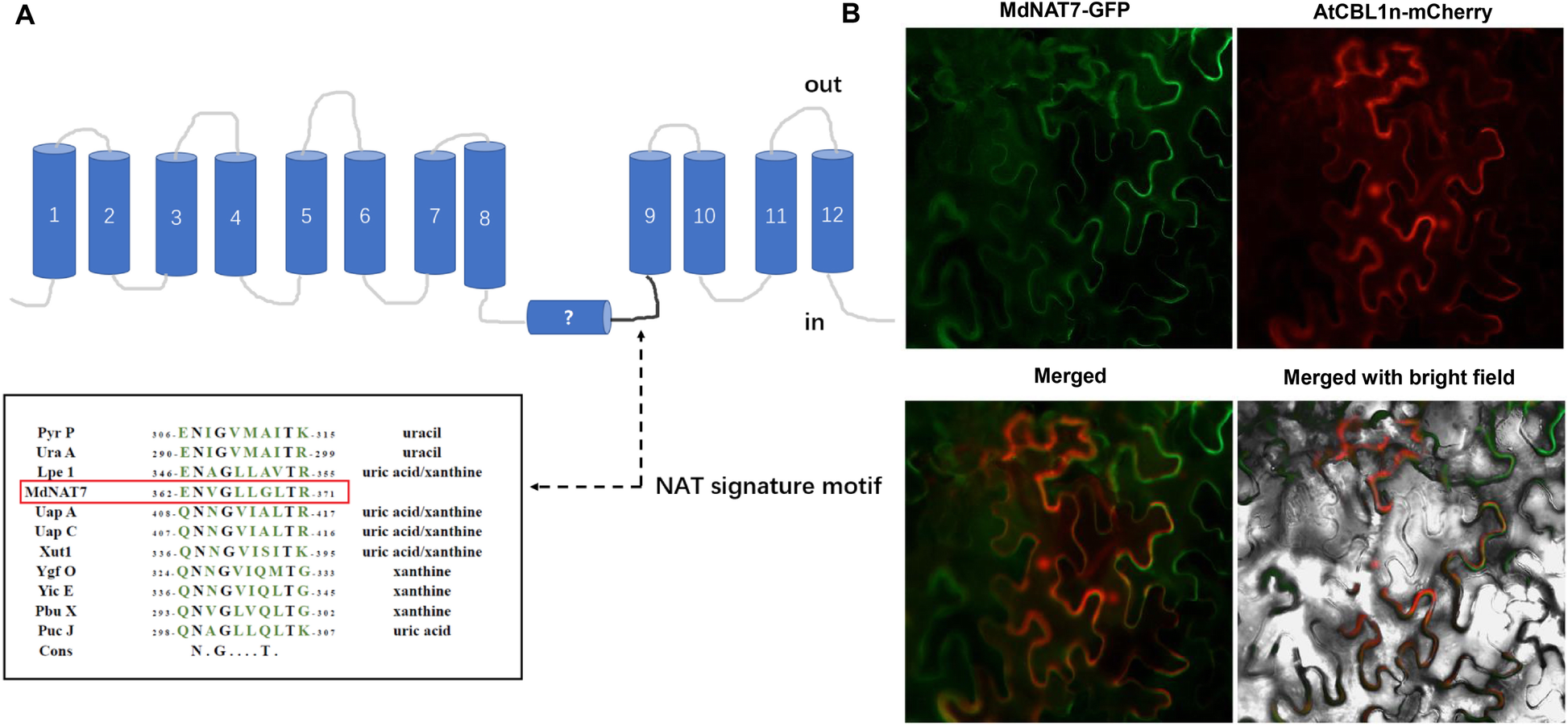
Fig. 1

Analysis and localization of MdNAT7.a.Model of the transmembrane structure of NAT proteins and the NAT signature motif. Pyr P,Bacillus subtilisPyr P (P31466), Ura A,Escherichia colihomologue Ura A (P0AGM7); Lpe 1, maize Leaf Permease 1 (NP_001150400.1); MdNAT7,M. domesticaNAT7 (MDP0000304285); Uap A,Aspergillus nidulansUap A (Q07307); Uap C,A. nidulansUap C (P48777); Xut1,Candida albicansXut1 (AAX22221.1); Ygf O,E. colihomologue YgfO (P67444); Yic E,E. colihomologue Yic E (P0AGM9); Pbu X,B. subtilisPbu X (P42086); Puc J,B. subtilisPbu J (O32139); Cons, Consensus refers to the nucleobase-ascorbate transporter motif.b.Localization of MdNAT7. The fusion protein of MdNAT7–GFP was transiently expressed in tobacco leaves and observed with confocal microscopy. MdNAT7–GFP fluorescence. The plasma membrane protein localization marker AtCBL1n-mCherry. Merged images. Merged with bright field. Scale bar = 50 μm
