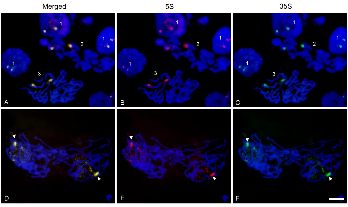
Figure 7

Fluorescencein situhybridization of 5 S (red) and 35 S rDNA (green) probes toHelichrysum bracteatumnuclei.Pictures in the first row(A, B & C)show rDNA signals in interphase: (field 1), metaphase chromosomes (field 2), and a prophasic cell (field 3). The second row(D, E & F)shows late anaphase/early telophase. One rDNA homolog is highly condensed (arrowheads) whereas the other is spread throughout the nucleus and decondensed. Bar – 10 μm.
