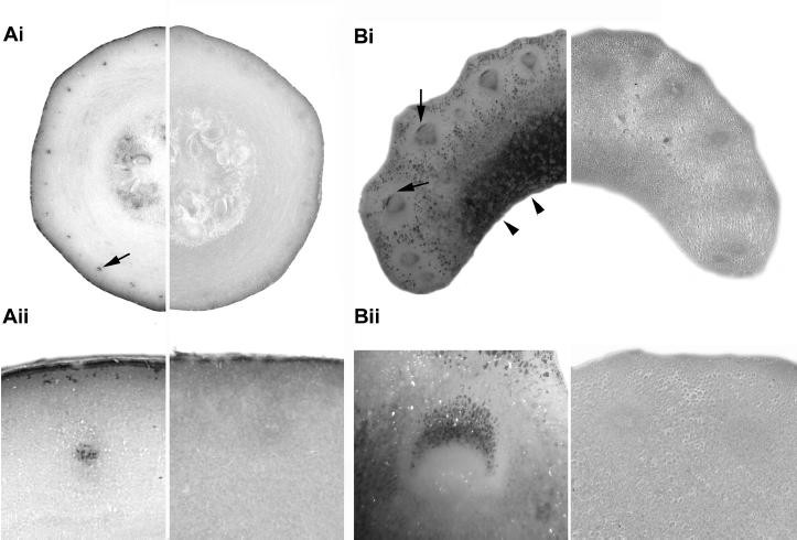
Figure 2

Localisation of AsA with methanolic AgNO3in courgette and celery tissues.The left side of each image shows sections stained immediately after excision and the right side control sections which were pre-incubated in 5% (w/v) CuSO4to oxidise endogenous AsA. Transverse section of courgette fruit (Ai) shows intense staining in the region of the outer vascular ring (arrows). Staining was also observed in the region surrounding developing seeds. Detail of the outer vascular area confirms that staining was confined to vascular regions (Aii). Staining was absent in control sections pre-incubated with 5% CuSO4。芹菜叶柄(Bi) intense staining was associated with vascular bundles (arrows) and storage parenchyma (tail-less arrows). Detail of the vascular region (Bii) shows intense staining associated with the phloem but not the xylem.
