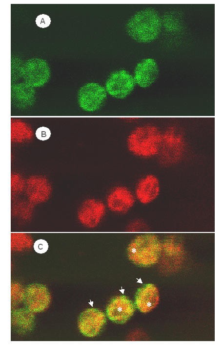
Figure 1

Confocal fluorescence micrographs of a pea leaf 24 h after biolistic transformation and transient expression of CAO-GFP fusion protein. (A), Location of the GFP tag is shown by green fluorescence. (B), Thylakoid membranes are indicated by the red fluorescence of Chl. (C), Overlay of green and red fluorescence signals. Green fluorescence of CAO-GFP was detected only within chloroplasts and was more prominent near the periphery of the organelles (arrows). Examples of regions of the chloroplast where Chl fluorescence predominated are shown byasterisks.
