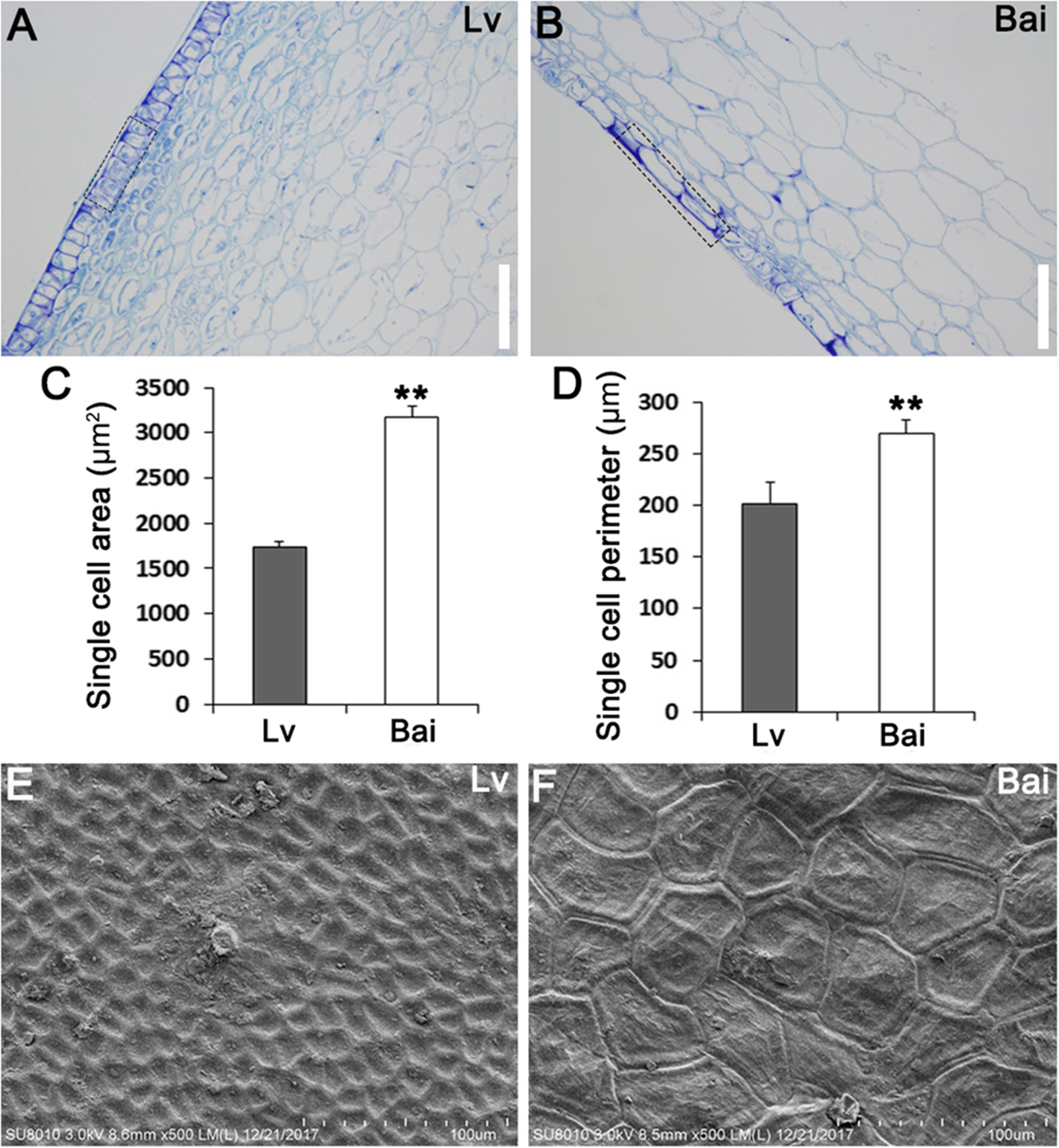
Fig. 3

Epidermal cells from Bai showed larger single cell area and perimeter.a,bObservation of paraffin section of fruit skins from Lv (a) and Bai (b).cSingle cell area of epidermal cells from Lv and Bai fruit skins.dSingle cell perimeter of epidermal cells from Lv and Bai fruit skin.e,fSEM observation of fruit skin from Lv (e) and Bai (f). Scar bar in (a,b): 150 μm. Data is presented as the mean ± standard deviation (n = 9). *0.01 ≤ P ≤ 0.05, **P ≤ 0.01, Student’sttest
