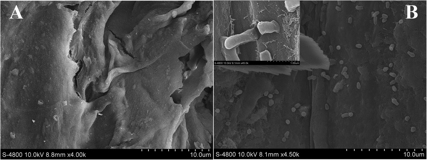
Fig. 1
From:Resistance analysis of cherry rootstock ‘CDR-1’ (Prunus mahaleb) to crown gall disease

Morphological observation of the wound surface viewed under a field emission scanning electron microscope.aImages of the wound in wounded control group at 5 days post-infection (dpi) at a magnification of × 4.00 k.bImages of the wound in treatment with inoculation at 5 dpi at a magnification of × 4.50 k
