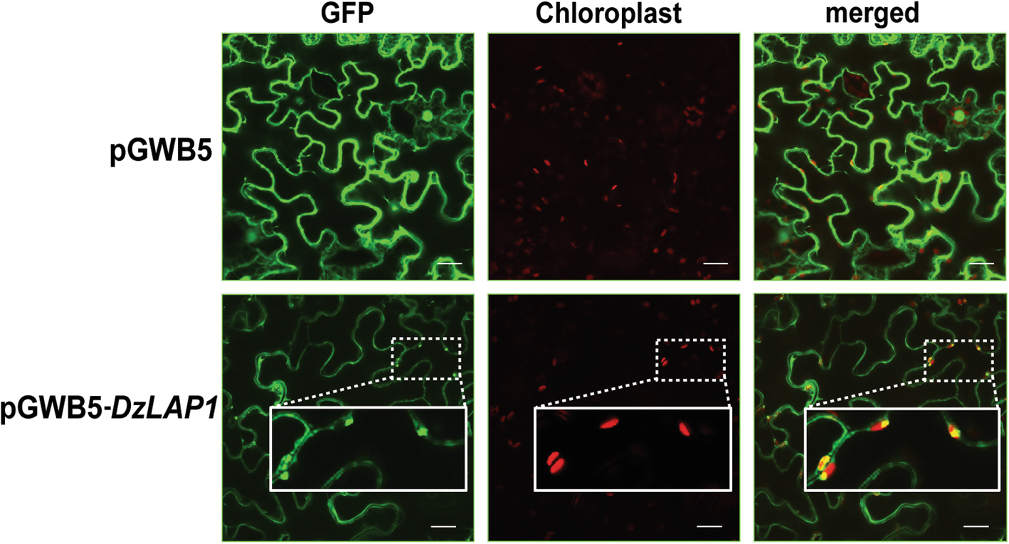
Fig. 6

Subcellular GFP-tagged DzLAP1 localisation in烟草benthamianaleaves. Confocal microscopic images ofN. benthamianaleaf epidermal cells infiltrated with pGWB5 (control; upper panel) and pGWB5-DzLAP1(lower panel). GFP fluorescence (GFP), chloroplast autofluorescence (chloroplast), and merged images are shown. Plastidial DzLAP1-GFP localisation is shown as enlargements in the insets. Scale bars: 20 μm
